Imagine your lab has decided to take the plunge and implement antinuclear antibody (ANA) testing in house, taking it off the send-out menu. You might first ask, What is the best method for ANA testing? Or, what if your lab already performs ANA testing, but the expert technologist who has been reading ANA indirect immunofluorescence (IIF) slides for 30 years has just announced that she is going to retire. This might prompt you to ask, Is it time for us to move from IIF ANA testing to a newer methodology?
These are important and relevant questions, but without easy answers. This review aims to provide practical information on ANA testing methodologies, including their diagnostic utility and performance characteristics.
ANAs refer to a collection of autoantibodies that target a variety of nuclear and cytoplasmic antigens. First described more than 50 years ago, ANAs remain the most sensitive serologic mark-er for evaluating patients with suspected connective tissue diseases (CTDs), also referred to as ANA-associated rheumatic diseases (AARDs) .
The diagnostic potential of ANAs originated with the discovery of LE cells, described as ma-ture polymorphonuclear leukocytes containing phagocytosed nuclear material. LE cells were so-named because they were found only in patients with systemic lupus erythematosus (SLE). LE cells could be produced in vitro by taking patient plasma and mixing it with peripheral blood from healthy controls that had been “damaged” by vortexing with glass beads.
Ultimately, research demonstrated that immunoglobulin from patient plasma was binding to nuclei from the “damaged” peripheral blood, which neutrophils in turn phagocytosed. IIF was used to further characterize this immunoglobulin, demonstrating its specific binding to cellular nuclear material. This immunoglobulin is what we now know as the ANA.
ANA testing generally involves two parts (2). First, for patients with a suspected AARD, a screening ANA is ordered to detect the ANA regardless of the antigen specificity. Second, for patients with positive screening assay results, additional tests characterize the antigen specificity of their ANA. Identifying the antigen specificity has important diagnostic and prognostic implications for patients. Although dozens of antigens have been associated with ANAs, only a small number are available for routine clinical testing. Depending on a patient’s clinical scenario, a positive ANA may require testing for anti-double standard DNA antibodies, antibodies against one or more of the extractable nuclear antigens (SS-A, SS-B, Sm, Scl-70, Jo-1, and RNP), anti-ribosomal P antibodies, or anti-centromere antibodies.
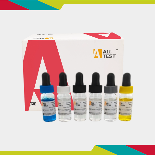
Three primary methods are available to clinical laboratories as screening ANA tests: IIF, enzyme immunoassay (EIA), and multiplex immunoassay (MIA) (Table 1) (3).
IIF detects antibodies that bind to a tissue substrate which, for ANAs, is usually fixed HEp-2 cells. IIF accomplishes this detection with a fluorescently labeled anti-human immunoglobulin. With EIA, an antigen mixture adhered to a solid surface (usually a 96-well plate) takes the place of the HEp-2 cells, and detection occurs through an enzyme-labeled anti-human immunoglobulin. MIAs are based on polystyrene bead sets distinguished from one another based on their fluorescent signature.
Each bead set is conjugated to a known ANA antigen, and the different sets are then combined into a bead cocktail. A patient sample is added to the bead cocktail, and binding of a patient antibody to any of the beads is accomplished with a fluorescently labeled anti-human immunoglobulin.Reporting of ANA Test Results
From a physician’s perspective, one of the most obvious differences between ANA screening methods is how results are reported. In most cases, MIAs are reported qualitatively as “ANA positive” or “ANA negative,” with screen results being based on the collective assessment of all the individual antigen specificities included in an assay. If all the included antigen specificities are negative, then the ANA screen is interpreted as negative. Conversely, if one or more of the beads show fluorescence exceeding a certain threshold, a sample would be identified as positive.
Importantly, for ANA positive samples, the identities of the antigen specificities are not revealed to the laboratory and thus are not reported to patients’ medical records. If a clinician wants to determine the antigen specificity of a patient’s ANA, he or she would need to order the clinically relevant tests.
In contrast, most EIAs are reported as a numeric value with an arbitrary unit of measurement. There is no traceable standard for these assays, so each manufacturer establishes the units and analytical measuring range for its tests. EIAs’ quantitation is based on light absorbance. The enzyme linked to the detection antibody converts a colorless substrate to a colored product, the absorbance of which is compared to a standard curve. Manufacturers will provide a recommended cutoff, which is the unit value above which a sample would be considered “ANA positive”.
As with MIAs, a positive EIA result does not reveal the antigen specificity of the ANA, and further testing would be necessary if a clinician wants to know those details.
ANA by IIF is generally reported with both a titer and a pattern. Labs screen all samples initially at a single dilution, usually 1:40 or 1:80. Any sample identified as positive at the screening dilution is titered out either to endpoint or to a pre-defined dilution, depending on the laboratory’s preference. The titer is determined by serial dilution, with the reported titer being the last dilution for which the IIF would be identified as positive. The pattern interpretation is based upon recognition of specific cellular features to which a patient’s antibody has bound (Figure 1).
Because IIF pattern interpretation is based on visual interpretation, standardization in reporting has been a challenge. The International Consensus on ANA Patterns (ICAP), a subcommittee of the Autoantibody Standardization Committee, promotes discussion and generates consensus regarding the morphologic features associated with specific ANA patterns (4). ICAP has also made recommendations regarding how laboratories should report ANA patterns. The group has defined six nuclear patterns as “Competent-Level”: homogeneous; speckled; dense fine speckled (DFS); centromere; discrete nuclear dots; and nucleolar.
ICAP recommends that any laboratory performing ANA by IIF should be able to accurately and reproducibly identify these patterns. The remaining nuclear patterns are designated as “Expert-Level” and might be recognizable only by individuals with particular expertise in IIF analysis.
When considering which ANA test to implement, understanding each method’s clinical sensitivity and specificity is critical. Many studies have compared the clinical sensitivity and specificity of the different methods. Because IIFs, EIAs, and MIAs report results so differently, these studies have focused primarily on qualitative agreement. Although seemingly very straight-forward, these types of comparisons are more difficult than they appear, largely because estimated sensitivities and specificities and the agreement between methods is heavily dependent on the cutoffs used to differentiate between positive and negative.
Historically, IIF has been considered the most sensitive method for identifying patients with AARDs. In a 2009 position statement on ANA testing methods, the American College of Rheumatology identified IIF as the “gold standard for ANA testing” primarily based on its high sensitivity (>95%) for the diagnosis of SLE (5). However, the statement also acknowledges that the specificity of ANA by IIF is a limitation.
In a cohort of patients for whom ANA testing was ordered as part of routine clinical care, we demonstrated that IIF at a titer cutoff of 1:40 had a sensitivity of 94% for the general diagnosis of AARDs (6). This was higher than the sensitivity of either EIA or MIA, at 74% and 67%, respectively. However, the IIF’s higher sensitivity was at the expense of specificity, which, at the 1:40 cutoff, was only 43%. In comparison, the corresponding EIA and MIA specificities were 80% and 87%, respectively. When we increased the cutoff for IIF to 1:80, the specificity improved to 62% but the sensitivity decreased to 84%.
Some data suggest that the titer of the ANA may help in distinguishing between patients with and without AARDs. In a study from 2011, Mariz et al. demonstrated that 45.8% of positive AN-As in healthy controls had a titer of 1:80, while 88.5% of ANA-positive AARD patients had an ANA titer ≥1:320 (7). Many laboratories that perform ANA by IIF are moving away from screen-ing at the 1:40 dilution, opting for improved specificity even with some loss in sensitivity. When labs use higher screening dilutions, the sensitivities of IIFs are on par with those of EIAs and MIAs. Although IIFs have the capability of maximizing sensitivity, from a practical perspective, EIAs and MIAs provide a good balance of sensitivity and specificity.
IIF’s sensitivity is attributed to its broad antigen specificity. This method detects antibodies against any of the hundreds of nuclear and cytoplasmic antigens present in a cell. However, not all antigen specificities are relevant for the diagnosis of AARDs. For example, the DFS pattern appears almost exclusively in patients with no evidence of an AARD (7). It has been suggested that the presence of the DFS pattern could be used to rule out an AARD in an individual with a positive ANA. The antigen specificity associated with this pattern has been identified as lens epithelial-derived growth factor, also referred to as DFS70 (8).
Further studies have con-firmed that monospecificity for DFS70 in the context of a DFS pattern is not consistent with an AARD. This pattern, and perhaps others like it that have yet to be characterized, may help to address some of the specificity challenges associated with ANA testing by IIF.
When labs are considering which ANA method to implement, availability of a qualified technologist to perform the testing is likely a significant concern. Other key considerations include throughput, workflow, and automation of a method.
Although automation of immunological testing has not reached the level of chemistry platforms, significant strides have been made over the last decade, particularly with EIAs and MIAs. EIAs can be performed manually, although more often than not, labs perform this testing on semi-automated or automated platforms. The semi-automated platforms may dilute patient samples and add reagents to the plate, but a technologist’s intervention might be required to wash and move the plate to an absorbance reader. A fully automated system processes an EIA in its entirety, only requiring technologists to load samples and reagents. Most MIA systems are also fully automated.
In addition, MIAs have the advantage of being random access, which facilitates improved workflows. In contrast, EIAs are batched, which, for labs with lower volumes of ANA orders, could have a negative impact on workflow and on turnaround times. Another advantage of MIA systems is they offer labs the opportunity to expand their test menus. Most MIA systems are not limited to ANA testing, and have reagents available for other autoimmune conditions (celiac disease, antiphospholipid syndrome, and vasculitis) and for infectious diseases (Epstein-Barr virus, HIV, and herpes simplex virus). Being able to perform additional testing and maximize an instrument’s utilization could make an MIA system an attractive option.
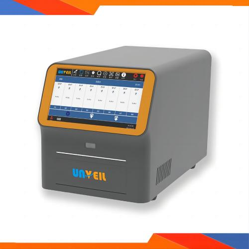
Historically, IIF has been the ANA method requiring the most clinical technologist resources and expertise, with automation limited to dilution of patient samples and perhaps addition of sample and reagents to slides. In addition, slide reading was a manual process that relied on experienced technologists to interpret numerous complex patterns.
Now, however, systems are available that automate almost the entire process, from slide processing to reading. Processing the slides includes not only sample and reagent pipetting but also slide incubation and washing. After processing, the slides can be moved to an enclosed microscope with a high-resolution digital camera, which obviates the need for a darkroom. This means such systems can be used on a bench in an open laboratory.
Cameras in these newer IIF systems capture several digital images from different areas of slides. The fluorescence intensity of the stain is measured, and values above a certain cutoff are considered positive. For samples identified as positive, the computer algorithm reads the pattern of and interprets the fluorescence intensities in the context of known ANA patterns. Although this step automates the previously manual process of slide reading, final qualitative and pattern interpretation still requires a technologist’s expertise. For each sample, a technologist must confirm the computer-generated result. If he or she disagrees, the result can be changed. Most automated readers recognize the common ANA patterns, and some identify certain mixed patterns.
More complex patterns unidentifiable by the computer still require a technologist’s interpretation. Some automated readers not only automate pattern interpretation at least partially but also estimate titers. These instruments use the fluorescence intensity of an image to estimate a sample’s titer rather than relying on serial dilutions. This can be accomplished either from a single patient dilution or a limited number of dilutions. As with pattern interpretation, an estimated titer can be replaced with a titer from serial dilutions, depending on the pattern and the technologist’s judgment.
Overall, although not completely automated by chemistry standards, the availability of automation for IIF, EIA, and MIA gives labs several options for complex ANA testing in a time of shrinking resources.
Over the last 10 years, ANA testing has experienced significant advances. Improvements in automation, development of new methods with better workflows, and even a clearer understanding of the diagnostic utility of this testing has widened the options for clinical laboratories.
However, choosing among EIA, MIA, and IFA is not easy, even when major guidelines are recommending IIF. No one-size-fits-all method exists, so each laboratory must make its own assessment as to which method is most beneficial for its patients and staff.


Tel:+86 -17280012527
Tel:+86-571-56267891

E-mail:info@alltests.com.cn

Add: 550 Yinhai street, Hangzhou Economic and Technologic Development Area 310018, P.R China
Copyright 2021 HANGZHOU ALLTEST BIOTECH CO.,LTD All Rights Reserved
浙ICP备16036562号-1
 浙公网安备 33011802001000号